
Conference Schedule

Day1: March 25, 2019
Keynote Forum

10:00-10:45
Biography
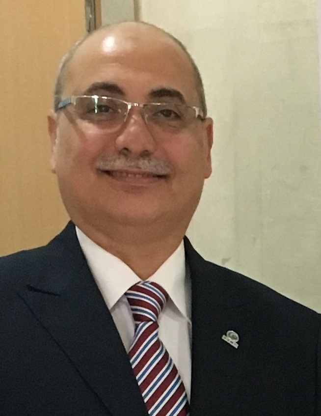
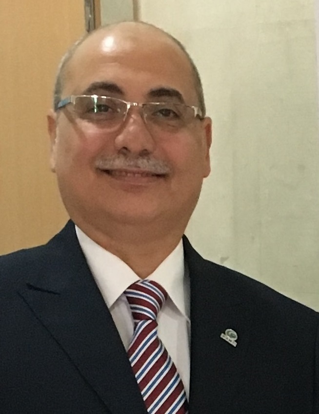
Dr. Sherif Abdelaal is Professor of conservative dentistry in faculty of oral and dental medicine, Cairo University, Egypt. He finished his PHD degree in Conservative Dentistry from the same faculty since 20 years ago. He has many published researches in local and regional journals in operative dentistry. He participated in many national and international conferences. He is now working as Consultant of Restorative Dentistry at King Abdulla Medical City Saudi Arabia
Abstract
Adhesion to dental substrate is the key foundation for modern esthetic dentistry that enables practitioners using of tooth colored direct and indirect resins and ceramic based restorations. At their foundation, direct anterior and posterior resin composites, endodontic posts, resin cores, and laboratory processed resin and ceramic veneers, inlays, onlays, and crowns all have bonding agents that function as the bridge between the restorative material or luting cement and tooth structure. To achieve clinical success with such restorations, good adhesion between restorative materials and tooth substrates is of crucial importance in order to ensure good marginal sealing, reinforcement of the tooth structure, and longer life of the restoration. During the last two decades, great advances in adhesive dentistry achieved forming what is called by “bonding revolution “in order to produce good adhesion to dental substrates. Modern adhesive systems seem superficially simple - merely a film to be painted on the tooth. Actually, they are often sophisticated and complicated chemical systems that have the challenge of sensitivity to the heroic oral environment where placed. So careful attention to appropriate use and good clinical technique is essential for getting the best performance out of the bonding system and, ultimately, the restoration. The presentation will address clinical manipulative tips for obtaining the highest performances, in terms of bond durability and stability of the adhesive interface using these adhesive systems starting from selecting the appropriate system, application tips, common manipulative errors and detecting possible failures.
10:45-11:30

10:45-11:30
Biography
Abstract
Objectives: To find out size of mastoid bone pneumatization (MBP) according to cleft lip and palate types of different severity and age subgroups, growing rate of MBP with aging , ear side dependence according to cleft side
Method: Measurement and analysis of mastoid bone pneumatization size on x-ray pictures (made according to Schuller technique) performed planimetrically using pixels separately for left and right ears. Study group include 14 bilateral (BCLP), 58 unilateral cleft lip and palate (UCLP) and 74 children with isolated cleft palate (ICP) (all groups have median age of 6.0years) suffered of recurrent episodes of otitis media with effusion (OME). Control group included 52 non-cleft children suffered of recurrent episodes of OME (median age of 6.0yr). Comparation of size of MBP of left vs. right ears for different age groups performed in different cleft lip and palate types.
Results: Size of MBP for total group of tested cleft palate ears was (7.3cm2), that is significantly smaller than MBP for total group of OME ears (8.12cm2) (p=0.0018). MBP in OME ears (8.12cm2) was bigger than in separate cleft type ears: BCLP (6,5cm2, p=0.0042), UCLP (7.4cm2, p=0.0052) and ICP (7,2cm2 p=0.0022). MBP in SOM ears showed faster rate of pneumatization growth (development) with aging(r=0.293, p=0.0035) than total group of cleft palate ears ( r=0.174,p=0.003). Mastoid bone pneumatization in BCLP and UCLP have smallest MBP size which do not grow significantlly with aging.MBP in isolated cleft palate have highest size if compared with other cleft types.This is only cleft tzpe with significant growing of MBP with aging.
Conclusion: Cleft types with highest severy of structural defects bilateral and unilateral cleft lip and palate have smalest MBP , and no tendency of growth in size with aging.Ears of children of isolated cleft palate type showed higher size and growing rate of MBP with aging than. High severity and structural defects in BCLP and UCLP are accompanied with retardation of development and growth of MBP, what makes them of high risk for further frequent infflamations and reccurence of OME,That contributes to retardation or/and slower development of mastoid pneumatization.
Tracks
- Orthodontics | Oral and Maxillofacial Surgery| Endodontics | Periodontics | Dental Implantology | Prosthodontics | Cosmetic Dentistry | Laser Dentistry | Oral Pathology
Location: Budapest, Hungary
Sherif Abdelaal
King Abdulla Medical City, Saudi Arabia
Chair
Jadranka Handzic
University Hospital Center, Croatia
Co Chair
11:50-12:20
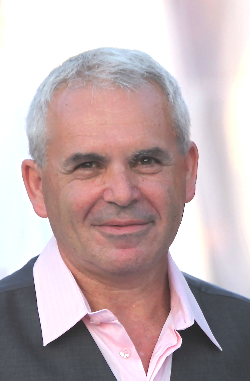
Uri Zilberman
Barzilai Medical University Center, Israel
Title: MIH and PEIR: Their effect on mineralization and treatment options

11:50-12:20
Biography
Abstract
Molar Incisor Hypomineralization (MIH) is caused by disturbance during the initial calcification and/or maturation of the enamel of the affected teeth. It happens during the first two years after birth, for permanent molars or during the third trimester of pregnancy for primary molars. The characteristic features of MIH: opaque stains that vary in tone between white, yellow and brown, post-eruptive enamel breakdown, atypical restorations and/or extensive caries with opacities at the margins, sensitivity, tenderness and difficulty to treat. In Pre-Eruptive Intracoronal Resorption (PEIR), currently of unknown aetiology- the dentine underlying the enamel crown of still unerupted teeth is progressively resorbed and replaced by soft granulomatous tissue leaving only a thin layer of unsupported enamel. The fragile enamel in such teeth fractures soon after eruption exposing the dental pulp to oral pathogens. In the absence of early intervention, the extent of destruction of the mineralized tissues resulting from PEIR, necessitates extraction in some 50% of affected teeth.
12:20-12:50

12:20-12:50
Biography
Abstract
Introduction Dental zirconia restorations present long-term clinical survival and be in service within the oral environment for many years. However, low temperature degradation could affect their mechanical properties and survival.
Objectives: Was to investigate the effect of two aging methods on the flexural strength and crystal structure of yttrium-stabilized zirconia (Y-TZP).
Material and methods: Thirty bar specimens were prepared from a yettria stabilized zirconia polycrystals and were divided into 3 groups (control, aged for 720,000 mechanical loads of 50N and 3600 thermal cycles, aged for 1 hour using autoclave). The aging procedures represent 3 years of clinical use. The specimens were loaded until fracture and the crystalline phase polymorphs of the material (tetragonal, t, and monoclinic, m, zirconia) were investigated by x-ray diffraction (XRD). Further investigations were done using scanning electron microscope (SEM). Data was statistically analysed using ANOVA test.
Results: Group B and C showed no statistical significance in their flexural strength with means of their break force (793.23±164.03) and (780.97 ± 257.25) respectively but statistically significant and higher than group A with mean (549.7 ± 54.14). The XRD showed nearly no change in the crystal structure between group A and B but an increase in the percent of monoclinic phase in group C. The SEM demonstrated a relatively homogenous size with particle size ranged between 400 to 570 µm for group A, while Group B and C showed an increase in particle size between 768 to 1150 µm respectively.
Conclusions: Both aging methods caused changes in the flexural strength and structure of the zirconia specimens with no significant difference between them.
13:50-14:20
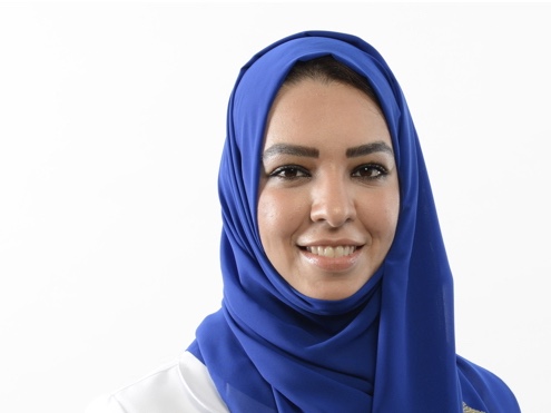
Ghadeer Islem Basunbul
King Abdul Aziz University, Saudi Arabia
Title: TMD Into perspectives: Evidence based diagnosis

13:50-14:20
Biography
Dr. Ghadeer Basunbul graduated with an honor degree and as the first on her class with a Baccalaureate in Dental Surgery (BDS) from King Abdul Aziz University, Faculty of Dentistry, Jeddah, Saudi Arabia in 2005. Between 2005 and 2007, Dr.G.Basunbul served as a demonstrator at the Department of Oral and Maxillofacial Rehabilitation at KAU. She obtained her Certificate in Advanced Graduate Studies in Prosthodontics (CAGS) and her Doctorate of Science in Dental Biomaterial and Restorative Sciences from Boston University, School of Dental Medicine in Boston, Massachusetts, USA in 2010 and 2013, respectively. Dr..Basunbul received Dr.David Baraban Excellence Award in Cosmetic, Prosthodontics and Implant Dentistry from Boston University, School of Dental Medicine in 2010. Since 2014, Dr. Ghadeer became a fellow of The Royal College of Dentists of Canada and joined the Oral and Maxillofacial Rehabilitation Department at King Abdul Aziz University, Faculty of Dentistry as a consultant and an assistant professor in prosthodontics. She participates in teaching and supervising the dental students at KAU as well as being engaged in teaching and certifying the Saudi Board Prosthodontic residents, Saudi Board Restorative residents and implant fellows. She participated in the establishment of Unident clinics in Jeddah city, KSA with a group of Consultants in all dental specialties where she also practices there. She participated in international meetings and has several publications in international journals in the field of dentistry. Her passion for education is encouraging her to move from a local lecturer in the field of prosthodontics and dental occlusion to an international speaker.
Abstract
TMD pain patients present dentists with challenges. For many, referral to experienced prosthodontists is the preferred action assuming that occlusion is involved. Providing therapeutic changes to the occlusion with appliances, re-creating muscle harmony, and prescribing diagnostic imaging to review the anatomy can provide invaluable information for the restorative dentist while providing relief and healing for most TMD patients. However, to provide predictable and successful results, the management of long term frustrated TMD pain patients starts with listening to consider when more than occlusion is involved. This requires knowledge and skills beyond restorative dentistry and occlusion. It requires evidence based comprehensive diagnosis. Understanding the etiology of temporomandibular disorders is fundamental for successful management. Yet there is much controversy and debate in this field. In this presentation, Dr.G.Basunbul discusses the long standing debate concerning the etiologic factors associated with TMD and where occlusion may fit. Understanding which etiologic factor(s) is predominant in the TMD is essential for helping their patients. Also this presentation presents an overview of orofacial pain and why it is often misdiagnosed and mismanaged. Often, our treatment strategies are based on our most familiar techniques and not always on the patient’s needs. Dentists understanding the global picture of orofacial pain is the start to reach proper diagnosis that leads to the most successful treatment strategy, including referral and ultimately improving the patient quality of life.

14:20-14:50
14:20-14:50
Biography
Abstract
Before any surgical intervention with the purpose of setting dental implants, the implantologist should not only do a general analysis of the case but first and most important thing is to do the detailed study of the region or area over which the intervention will take place. So, the implant-prosthetic rehabilitation requires that alveolar crusts must have enough bone in good quality and on which we can work. But this is not always possible. Different factors influence this inadequacy which in some cases is an obstacle that should be treated seriously by the physician.
Purpose: The main focus of this paper is the detailed analysis of the factors causing bone atrophy and not only the specification of it according to the jaws but also the detailed study by specific regions as well. Alternative methods to overcome obstacles whether or not surgical will not be treated only theoretically but will also be illustrated with clinical cases that have been performed after patients' consensus. Dental elements loss causes artificial shortage of biomechanical bone stimulation, which is of utmost importance in maintaining its physiological volume. Such loss results in bone absorption which may be hidden in the first 6 months, but then it continues with a bone loss of about 0.1 mm per year.
In the upper jaw, the absence of teeth causes the reduction of the bone thickness between the maxillary sinus and the alveolar ridge up to 1mm. In such situations, the maxillary sinus pressure pushes the bone floor down. By the other hand, the present inflammation causes a generalized or localized thickening of the sinus membrane. Surgical maxillary sinus floor elevating is one the pre-implant reconstructive surgical techniques. When this technique is correctly done it does not significantly affect the physiology of maxillary sinus.One of the auxiliary ways for implant placement in the alveolar ridges with horizontal dimension loss is the osteotomy of the alveolar process (split ridge). The alveolar split ridge method has advantages over the grafting, because it significantly reduces the time of the treatment. Also bone compression and increasing trabecular density are other advantages of this technique. Intercortical space filling is done with autograft, allograft or xenograft. In generally, these surgical auxiliary techniques are used during interventions for atrophic bone implantation.
Material and Method: In this paper I have used my personal theoretical-practical experience in the surgical treatment of patients who had undergone atrophic bone surgery for a period of several years. In this paper we will present some clinical cases treated at our clinic. Clinica case:The MS 50-year-old patient appears in our clinic after a failed implant surgery on both jaws. The panoramic graph gives this view. It is snapped into the upper jaw and after its prosthesis the control chart gives this view. The patient rightly needs to replenish the lower jaw. Bone atrophy is expressed both in length and width. There is a correctional intervention that is performed using the autologous bone graft, while the beneficial membrane is also used from enriched plasma.
Results: The results of the interventions were not only positive but the success limits were equal to the placement of simple implants. We cannot fail to mention that in all cases horizontal augmentation and post-stratigraphic implant management is accomplished through directed regeneration of the bone (GBR - guided bone regeneration). The main purpose of these procedures is to ensure long-term implant sustainability through successful and predictable bone regeneration with as few complications and additional surgical stages as possible.
Conclusion: The bone volume determines the individual evaluation criteria for the selection of the reconstructive technique that influences since in the beginning the selection of the best procedure for the morpho-functional restoring of soft and hard tissues and having the utmost care in aesthetics of implant prosthetics. The available techniques in implants surgeon hands, to obtain enough bone volume for implants’ positioning, are numerous and often sophisticated. Their implementing in implant surgery is the main duty of the doctor who takes over the surgical treatment of the patient.
14:50-15:20
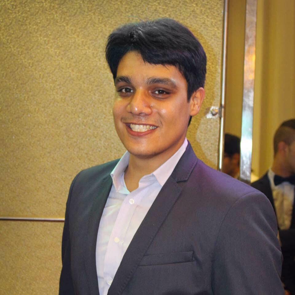
Omar Tarek Farouk
Delta University for Science and Technology, Egypt
Title: Smile makeover full protocol

14:50-15:20
Biography
Dr. Omar Tarek Farouk has completed his bachelor degree at the age of 25 years from Mansoura University and Master Degree of fixed prosthodontics from Ain Shams University concerning about aesthetic dentistry. He work as a clinical demonstrator at faculty of dentistry Delta University for science and technology. He attended more than 9 international conferences in his country, as a speaker. Has been serving as a director in 13 smile makeover courses in Egypt.
Abstract
Nowadays, as we entering era of adhesion, Side by side with increasing aesthetic demand between our patients. It is very important to meet our patient expectations. Smile makeover is a multifactorial high challengeable treatment option. Through this lecture we will see the different stages to produce proper smile makeover, clinical tips and tricks of each stage. Easy and efficient protocol for smile designing. Types of veneer preparation and how to select proper preparation for different case scenarios. How to manage complications. Type of ceramics used in veneers. Different cementation and adhesion protocols. Systematic approach for cementations. Different case scenarios.

15:20-15:50
15:20-15:50
Biography
Abstract

16:10-16:40
Biography
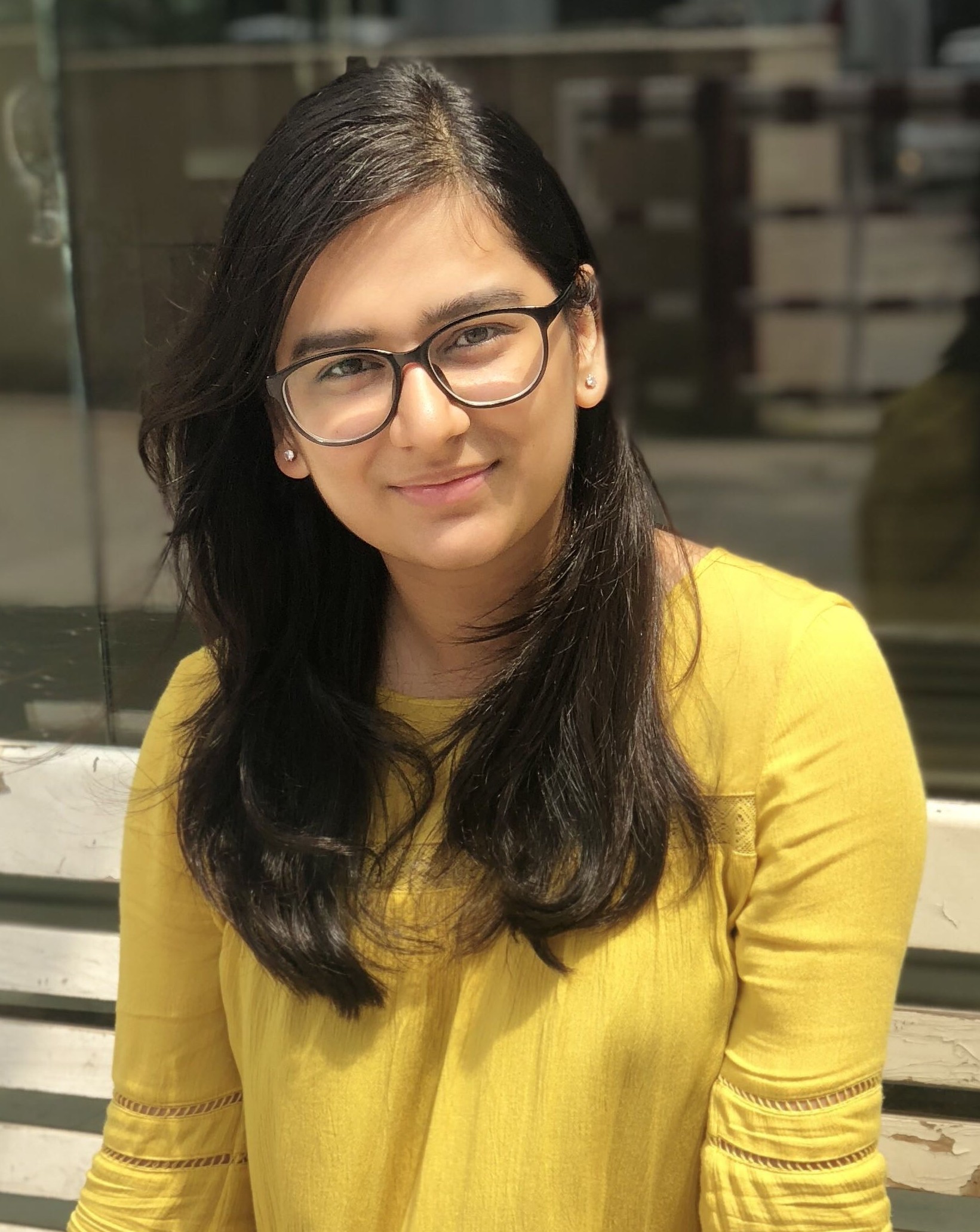
Arshdeep Kaur is pursuing Bachelor of Dental Surgery and currently doing her internship at a reputed college and hospital Dr Harvansh Singh Judge Institute of Dental Sciences and Hospital, Panjab University, Chandigarh, India. She will be completing her internship in July,2019.
Abstract
Edentulism has been a serious public health problem in industrialized countries due to population ageing and in developing countries due to poor oral care. The life quality and nutrition intake are impacted for edentulous patients. Historically, the complete removable denture is the last prosthetic procedure to switch to digital techniques.
Computer-aided design and Computer-aided manufacturing (CAD/CAM) has emerged as a new approach for the design and fabrication of complete dentures. However, unlike the extensive use of this new technology in other aspects of dentistry, the use of CAD/CAM was limited in the production of complete dentures due to lack of CAD software until recently. Several systems are now available including the Wieland Digital Denture which offers a complete procedure.
Virtual complete dentures have been successfully designed using the software through several steps including generation of 3D digital edentulous models, model analysis, arrangement of artificial teeth, trimming of relief area and occlusal adjustment.
The practitioner’s role has been simplified. Removable denture design has used the same classical procedures for more than fifty years despite their being associated with many risks of errors and long laboratory and clinical procedures. Thus the advantages of setting up a digital chain seemed obvious.

16:40-17:10
Tanvi Samujh
Panjab University, India
Title: Autotransplantation: An evolving treatment modality in modern dentistrypatients

16:40-17:10
Biography
Tanvi Samujh has passed Bachelor of Dental Surgery fourth year exam from Dr. Harvansh Singh Judge Institute of Dental Sciences & Hospital (HSJIDS) Panjab University, Sector 25, Chandigarh (U.T.) - India in June 2018 and is currently pursuing Internship in the same Institute.She had attended the 29th Annual World Congress on Dental Medicine & Dentistry at New York (USA) from October 16-18, 2017 and was awarded Best poster award for her presentation along with the moderator certificate. Apart from this she has attended 3 national conferences and presented posters.
Abstract
The procedure of transplantation of teeth from one individual to another has long been discussed in texts as a possible procedure for replacement of teeth. It, however, didn’t gain popularity on grounds of ethical issues and was also seen to be accompanied with failures like tissue rejection and disease transmission.
To overcome the above mentioned failures ,the procedure of autotransplantation came into view.
Autotransplantation is defined as the surgical movement of a tooth from one position to another , within the same person. Patient age, donor tooth position, extra oral time , recipient site and developmental status of donor tooth are some of the prognostic factors evaluated during the study. Congenitally absent tooth , loss of maxillary incisors due to trauma are few of the many indications of autotransplantation. Advantages include bone induction, preservation of Periodontal ligaments, normal eruption of tooth.
Autotransplantation is ,thus, evolving as an treatment modality in modern dentistry.


17:10-17:40
Biography
Anisha is pursuing Bachelors of Dental Surgery and currently doing her internship at a reputed college and hospital Dr. Harvansh Singh Judge Institute of Dental Sciences and Hospital, Panjab University, Chandigarh, India. She will be completing her internship in July, 2019.
Abstract
Pineapple (Ananas cosmosus) is a tropical plant belonging to the family of Bromeliacea. It has been used as folk medicine by the natives of the tropics. It contains Vitamin C, Vitamin A, Beta-carotene, calcium, manganese, folate, potassium, thiamine and is the only natural source of a complex of enzymes called bromelain.
Though the exact chemical structure of all active components of bromelain is not fully determined, this substance has shown distinct pharmacological promise. Its properties include: interference with growth of malignant cells, inhibition of platelet aggregation, fibrinolytic activity, anti-inflammatory action.
These biological functions of bromelain, a non-toxic compound, have therapeutic values in modulating: tumour growth, blood coagulation, inflammatory changes. The mechanism of action of bromelain affecting these varied biological effects relates in part to its modulation of the arachidonate cascade.
A hearty portion of pineapple may return stained teeth to their shiny selves as bromelain acts as a natural stain remover. Also pineapple has an incredible ability to break up plaque. In third molar surgery and placement of implants, consumption of pineapple and pineapple juice two days prior to surgery and two days after surgery helps prevent swelling and bruising. This is because of the proteolytic action of bromelain that counteracts inflammation. These varied benefits of pineapple presents it as a wonder fruit that acts as a natural cleaner and healer for our general health as well as of our thirty-two pearly whites.
17:40-18:10
Tanvi Thakur
Panjab University, India
Title: Green dentistry: A paradigm shift towards sustainability

17:40-18:10
Biography
Tanvi Thakur is pursuing Bachelors of Dental Surgery and currently doing her internship at a reputed college and hospital Dr. Harvansh Singh Judge Institute of Dental Sciences and Hospital, Panjab University, Chandigarh, India. She will be completing her internship in July, 2019.
Abstract
In the contemporary world of scientific advancements and discovery, sustainability and eco-compliance have been the driving forces for all new technologies across all fields. Many industries have shown paradigm shifts in established processes and methodologies to move towards sustainable and environment friendly future.
Amidst this changing world scenario, Green dentistry comes as a promising advancement in mitigating the ill-effects of dental wastes on the environment, and as an upgrade to the existing dental practices which are energy and resource intensive.
Green Dentistry is a high-tech approach that reduces the environmental impact of dental practices and encompasses a service model for dentistry that supports and maintains wellness. In essence, it encompasses the long established concept of ‘4R’ – Reduce, Reuse, Recycle and Rethink. In today’s world of dying whales and vanishing forests, it becomes a professional obligation and a social responsibility for the dental community to adopt greener measures and play our part in environment protection.
It’s time to change colour coded bags to GREEN!